
Facial skin cancer most often manifests as a skin lesion that does not heal, changes shape or color over several weeks. The face has a high cumulative sun exposure, making it particularly affected by basal cell and squamous cell carcinomas. Recognizing these lesions with the naked eye remains a delicate exercise, as their appearance varies according to the type of tumor, skin tone, and stage of development.
Why a photo of a facial skin lesion can be misleading
Most online content offers photo galleries to help spot skin cancer. These images are useful as a starting point, but they come with biases that are rarely mentioned.
Research published in Frontiers in Medicine in 2026 on dermatological photography shows that lighting, angle, and resolution significantly alter the appearance of a lesion. The same spot photographed under direct natural light or artificial lighting may appear flat in one case and raised in another. On the face, the contours of the nose, cheekbones, and arches create shadows that enhance or obscure the outlines of a lesion.
An analysis of the British healthcare system in 2026 revealed that nearly half of the initial photos submitted for algorithmic analysis were not usable. Areas hidden by hair, lack of sharpness, or inadequate framing account for most rejections.
Comparing a photo taken with a phone in a bathroom to a standardized clinical image often leads to false conclusions. To view documented images of facial skin cancer in a medical setting, a specialized reference remains more reliable than a generic image search.
Basal cell carcinoma on the face: distinctive signs
The basal cell carcinoma (BCC) is the most common form of skin cancer. It preferentially develops in areas chronically exposed to the sun: nose, eyelids, forehead, temples.
On the face, BCC often appears as a small, pearly, translucent nodule, sometimes with fine visible blood vessels (telangiectasias). The lesion may also present as a slightly raised pink plaque or a wound that intermittently bleeds and then seems to heal before reopening.
What distinguishes BCC from a pimple or a scar
An inflammatory pimple evolves over a few days. A BCC persists for several weeks without fully healing. The lesion is not painful, may not itch, and progresses slowly. It is precisely this discretion that delays consultation.
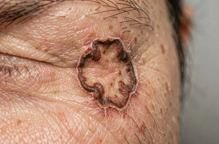
Another characteristic: an ulcerated BCC presents a central crater surrounded by a shiny peripheral rim. This morphology is very characteristic, but it only appears at a locally advanced stage.
Squamous cell carcinoma of the face: a more aggressive lesion
The squamous cell carcinoma (SCC) often develops on a background of actinic keratosis, those small rough and dry patches that can be felt before they are seen. On the face, the lips (especially the lower lip) and ears are common locations.
The typical lesion resembles a thick, firm crust, sometimes shaped like a skin horn, that does not fall off on its own. Unlike BCC, squamous cell carcinoma can grow rapidly over a few weeks and become painful to the touch. It presents a risk of metastasis, making it more concerning than basal cell carcinoma when diagnosed late.
- A persistent crust on the lip or ear that returns after each scratching attempt warrants prompt dermatological advice.
- A firm, pink to red nodule that visibly enlarges over a few weeks, especially in a person with a history of intense sun exposure.
- A scaly and hardened plaque on the forehead or temple, resistant to moisturizing creams, that does not improve after a month.
Facial melanoma: the ABCDE rule and its limitations
Melanoma remains the most dangerous skin cancer, although it is less common than carcinomas. On the face, it can appear on a pre-existing mole or arise de novo on seemingly healthy skin.
The ABCDE rule is a widely used visual screening tool:
- Asymmetry: the lesion is not identical on both sides of a central axis.
- Borders irregular: jagged, poorly defined, or notched contours.
- Color heterogeneous: presence of multiple shades (brown, black, red, white, blue) within the same lesion.
- Diameter: a lesion larger than a pencil eraser draws attention, but smaller melanomas exist.
- Evolution: any recent change in size, shape, color, or thickness.
What the ABCDE rule does not cover on the face
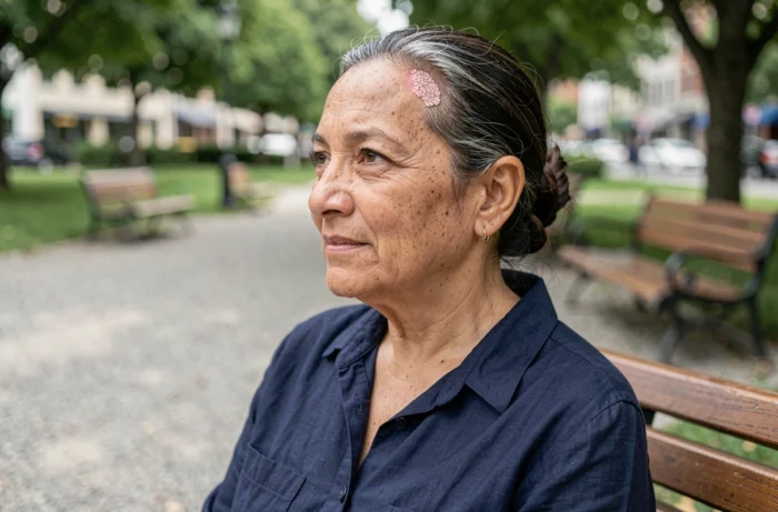
Some facial melanomas, particularly Dubreuilh melanoma (lentigo maligna), progress very slowly as an irregular brown spot, often confused with a age spot. This confusion is common among people over 60 who have been exposed to the sun for decades.
Recently tested artificial intelligence tools for skin screening also show limitations. The NHS analysis in 2026 highlighted that a significant number of carcinomas would have been missed if the decision had relied solely on the algorithm, without review by a dermatologist. AI does not replace clinical examination with a dermatoscope.
When to consult a dermatologist for a facial lesion
Any facial lesion that does not heal after three weeks, bleeds for no apparent reason, or changes color justifies a dermatological consultation. Self-monitoring remains a useful first filter, but a photo alone, taken outside a medical setting, does not allow for a diagnosis.
A dermatologist uses a dermatoscope (polarized magnifying glass) that reveals structures invisible to the naked eye and in a standard photo. It is this examination, complemented if necessary by a biopsy, that confirms or excludes skin cancer. Early detection of a carcinoma or melanoma of the face significantly improves treatment options and reduces the risk of aesthetic sequelae related to surgery.